HOME > Research Summaries > Novel Fluorescence Imaging Techniques with Functional Nano-particles for Medical Application
Research Summaries

Novel Fluorescence Imaging Techniques with Functional Nano-particles for Medical Application
Noriaki Ohuchi
Professor, Department of Surgical Oncology, Graduate School of Medicine, Tohoku University
E-mail:![]()
Abstract
Nanomedicine is the application of nanotechnology to prevention, diagnosis and treatment of human disease. It has a potential to change medical science dramatically in the 21st century. However, the research field is in its infancy, and it is necessary to grasp mechanism of pharmacokinetics, the toxicity on the occasion of application to medical treatment, in particular on the aspect of safety of the materials and devices. Here, we describe generation of CdSe nanoparticles, Quantum Dots (QDs) conjugated with monoclonal anti-HER2 antibody, Trastuzumab, for molecular imaging of breast cancer cells. The QDs-Trastuzumab complex coated with PEG was successfully made without decreasing the titer of antibody. We established a high resolution of 3D in vivo microscopic system as a novel imaging method at molecular level. The cancer cells expressing HER2 protein were visualized by the nanoparticles in vivo at subcellular resolution, suggesting future utilization of the system in medical applications including drug delivery system to target the primary and metastatic tumors. We also describe fluorescent tomonography by acousto-optic imaging with fluorescent nono-particles for sentinel node navigation for breast cancer surgery in experimental model, which have shown the potential to be an alternative to existing tracers in the detection of the sentinel nodes in depth.
Future innovation in cancer imaging, not only at cellular level but also at molecular level, by synthesizing diagnostic agents with nanoparticles, is now expected.
1. Introduction
Real-time single particle tracking using quantum dots (Qdots) has been applied to the study of drug delivery. Qdots fluorescence nanocrystals, were thought to be as the marker because of their intense brightness and stability, in contrast to organic dyes. In cultured cells, single particle tracking has yielded invaluable information on the function of purified proteins. Recent work shows the antibody-conjugated Qdots have allowed real-time tracking of single receptor molecules on the surface of live cells. However, no real-time single particle tracking in live animals have been reported, and it is uncertain that single particle of Qdots could be observed and tracked in live animals.
Treatment for cancer with minimum invasive surgery without lymph nodes dissection based on sentinel lymph node (SN) navigation surgery has become a major concern on the aspects of made-to-order and low-invasiveness medicine. Several radioisotopes and dyes are utilized for SN detection in standard methods, however, each detection method has advantages and disadvantages. To make up for the disadvantages, we aimed at developing a new non-invasive method using fluorescent beads of uniform nano-size that could efficiently visualize SN from outside the body and we have determined the appropriate size and fluorescent wavelength. On the other hand, there is an another problem in fluorescence measurement. The fluorescence measurement has a limitation for depth. In this study, we tried to develop a novel technique based on acousto-optic imaging.
These methods should aid the adoption of a fluorescence measurement method in the future.
2. Methods
2.1. Single Molecular Tracking
Qdot was conjugated to the monoclonal antibody trastuzumab (QT complex). The QT complex was injected intravenously into mice implanted with HER2 expressing tumor (KPL-4). We analyzed the movement of single functional Qdots in tumors of mice from a capillary vessel to cancer cells. To observe single Qdot particles in tumor tissue, we used a dorsal skin fold chamber (DSFC) model.

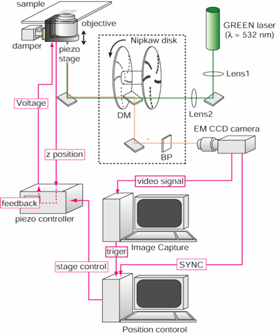
Fig.1. The 3-D intravital imaging system for visualization of QT-complex in mice.
The imaging system, consists of confocal scanner unit employing a Nipkow type disc and the electron multiplying charge coupled device camera, facilitates high resolution in vivo single particle tracking at a video rate with spatial resolution of 30 nm.
Quantitative and qualitative information such as velocity, directionality and transport mode was obtained using time-resolved trajectories of particles.
2.2. Acousto-Optic Modulation Imaging
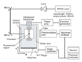
The experimental setup was comprised of a continuous wave diode pumped solid state laser system as a light source, an ultrasound transducer, and a photomultiplier tube (PMT) as a detector (Fig. 2). A tissue phantom is suspended independently from the holder, which is located above the water tank; the phantom is immersed in the water without touching the water tank (Fig. 2).
The imaging experiments are conducted using a wavelength of 532 nm and an incident beam signal intensity of 60 mW.

Fig. 2. Schematic of the experimental setup.
3. Results
3.1. Single Molecular Tracking
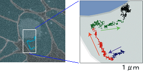
Two hours after the injection, many complexes had migrated into the tumor interstitial area close to the tumor vessels. Six hours after the injection, QT-complexes had bound to the KPL-4 cell membrane on which the HER2 protein is located. We also observed in pursuing the transport of QT-complexes from the peripheral region of the cell to the perinuclear region (Fig.3).
3.2. Acousto-Optic Modulation Imaging
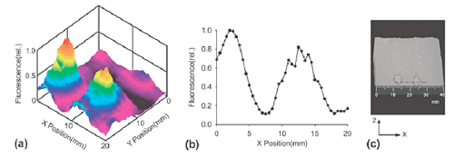
We could observe two fluorescent regions in a phantom with an embedded 9 mm gap along the X axis. Both the image Fig. 4(a) and the X-axis profile Fig. 4(b) show the two well-separated fluorescence peaks, which correspond to the embedded positions of both regions, as shown in the photograph sectioned on the longitudinal plane of the phantom Fig. 4(c).
4. Conclusion
This approach thus should afford great potential new insight into particle behavior in complex biological environments. Nanocrystal semiconductor quantum dots conjugated with antibody may serve fundamentally as new materials controllable for medical purposes including cancer molecular imaging.

Fig. 3 Tracking image of a QT-complex in a cancer cell.

Fig. 4. Tomographic image of fluorescence observed with a phantom.
References
[1] Takeda M, Kobayashi M, Takayama M, Suzuki S, Ishida T, Ohnuki K, Moriya T and Ohuchi N. Biophoton detection as a novel technique for cancer imaging. Cancer Science 95 (8), 656-661, 2004
[2] Kobayashi Y, Misawa K, Kobayashi M, Takeda M, Konno M, Satake M, Kawazoe Y, Ohuchi N, Kasuya A. Slica-coating of fluorescent polystyrene microspheres by a seeded polymerization technique and their photo-bleaching property. Colloids and surfaces A: Physicochem. Eng. Aspects 242, 47-52, 2004.
[3] Kobayashi Y, Misawa K, Takeda M, Kobayashi M, Satake M, Kawazoe Y, Ohuchi N, Kasuya A, Konno M. Slica-coating of AgI semiconductor nanoparticles. Colloids and Surfaces A: Physiochemical and Engineering Aspects, 251: 197-201, 2004
[4] Kasuya A, Sivamohan R, Barnakov YA, Dmitruk I M, Nirasawa T, Romanyuk VR, Kumar V, Mamykin SV, Tohji K, Jeyadevan B, Shinoda K, Kudo T, Terasaki O, Liu Z, Belosludov RV, Sundararajan V, Kawazoe Y. Ultra-stable nanoparticles of CdSe revealed from mass spectrometry. Nature Materials 3, 99-102, 2004.
[5] Nakajima M, Takeda M,Kobayashi M, Suzuki S, Ohuchi N. Nano-sized fluorescent particle as a new tracer for sentinel node detection: An experimental model for decision of appropriate size and wavelength. Cancer Science 96 (6), 353-356, 2005.
[6] Zhou X, Kobayashi Y, Ohuchi N, Takeda M and Kasuya A, Strong luminscence of CdSe nanoparticles by surface modification with gadmium (II) hydrous oxide. International Journal of Modern Physics B 19, 2835-2840, 2005.
[7] Zhou X, Kobayashi Y, Romanyuk V, Ohuchi N, Takeda M, Tsunekawa S and Kasuya A, Preparation of silica encapsulated CdSe quantum dots in aqueous solution with the improved optical properties. Applied Surface Science 242, 281-286, 2005
[8] Kasuya A, Noda Y, Dmitruk I, Romanyuk V, Tohji K, Kumar V, Belosludov R, Kawazoe Y, Ohuchi N. Stoichiometric and ultra-stable nanoparticles of II-IV compound semiconductors. Eur Physical Journal D 34, 39-41, 2005.
[9] Kobayashi Y, Misawa K, Kobayashi M, Takeda M, Konno M, Satake M, Kawazoe Y, Ohuchi N, Kasuya A, Silica-coating of fluorescent polystyrene microspheres by a modified Stober method and their stability against photo-bleaching. e-Polymers, no. 052, 1-8, 2005.
[10] Li-Shishido S, Watanabe TM, Tada H, Higuchi H, Ohuchi N. Reduction in nonfluorescence state of quantum dots on an immunofluorescence staining. Biochem Biophy Res Com 351 (1), 7-13, 2006
[11] Tada H, Watanabe T, Higuchi H, Ohuchi N. In vivo real-time tracking of single quantum dots conjugated with monoclonal anti-HER2 antibody in tumors of mice. Cancer Res 67 (3), 1138-44, 2007.
[12] Park YS, Liz-Marzán LM, Kasuya A, Kobayashi Y, Nagao D, Konno M, Mamykin S, Dmytruk A, Takeda M and Ohuchi N, X-ray Absorption of the Gold Nanoparticles with Thin Silica Shell. J Nanosci Nanotechnol 6 (11), 3503-06, 2006.
[13] Mamykin S, Kasuya A, Dmytruk A, Ohuchi N. Photocurrent of nanoassembled Si film in contact with electrolyte. J Alloys Compounds 434-435, 718-720. 2007.
[14] Park YS, Kasuya A, Dmytruk A, Yasuto N, Takeda M, Ohuchi N, Sato Y, Tohji, K Uo M, Watari F. Concentrated colloids of silica-encapsulated gold nanoparticles: colloidal stability, cytotoxicity, and X-ray absorption. J. Nanosci. Nanotech. 2007 (in press).
[15] Park YS, Dmytruk A, Dmitruk I, Noda Y, Kasuya A, Ohuchi N. Aqueous-phase synthesis of ultra-stable small CdSe nanoparticles. J. Nanosci. Nanotech. 2007 (in press)