HOME > Research Summaries > Normal Brain Aging and Its Risk Factors-Analyses of Brain MRI Database of 2,000 Healthy Japanese Subjects
Research Summaries

Normal Brain Aging and Its Risk Factors-Analyses of Brain MRI Database of 2,000 Healthy Japanese Subjects
Hiroshi Fukuda
Professor
Department of Nuclear Medicine and Radiology, Institute of Development, Aging and Cancer
E-mail:![]()
1. Introduction
Recent developments in imaging modalities are remarkable. We can visualize precise brain morphology and function by X-CT, MRI, PET, SPECT, fMRI and NIRS. Nowadays, brain researches using these techniques play an important role in neuroscience for living humans.
Most remarkable recently developed method for image analysis is a voxel based morphometry (VBM). It includes anatomical standardization of the brain to a standard brain, brain tissue classification, and finally voxel based statistical analysis based on general linear model. This technique enables us to extract brain regions which show correlations between tissue volume and variables, such as age, sex and subject’s characteristics. We can analyze not only age-related normal changes but also diseased brain, such as dementia and schizophrenia. It has been believed that functional imaging precede structural imaging to detect early pathological findings of the diseases. However, recent development of high resolution structural imaging and sophisticated analytical technique enable us to detect the brain disease at very early stage.
We have collected 2,000 brain MRI of healthy Japanese and constructed an MRI database together with their characteristics such as age, sex, blood pressure, present and past disease history and cognitive functions [1]. This is a largest database in Japan and one of the largest one in the world. We have been analyzing these data and obtained the following results until now.
(1) We measured volume of each brain tissue segment separately. Gray matter volume linearly decreased with age, while white matter volume remained unchanged during aging. CSF volume increased with age [2].
(2) We calculated regression line for the gray matter decrease with age (Fig. 1). Then, we defined “a normally aged brain for his/her age” as the brain which has gray matter volume on the regression line (this is a first objective criteria of normally aged brain). Figure 2 showed a normally aged brain and atrophic brain for 70 years old.
(3) We analyzed correlation between regional gray matter volume and subject’s characteristics. We found that gray matter volume negatively correlated with systolic blood pressure2) and alcohol drinking [3].
(4) We analyzed correlations between gray matter volume and mental status of the subjects and found that subthrshould depression, which was tested by geriatric depression scale (GDS) had smaller volume of gray matter in medial frontal lobe than that in healthy subjects [4].
(5) White matter hyperintensities on T2-weithted MRI increased with age and is considered as ischemic changes. However, their pathological significance remained unknown. To clarify pathological significance of the lesion, we developed an algorism for automatic detection and quantification of the lesion.
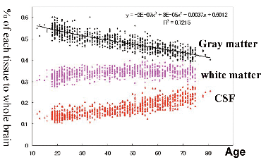
Fig. 1. Age-related volume change of the human brain
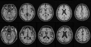
Fig. 2. T1 weighted brain MRI of 70 year-old male
Upper: a normally aged brain for his age. Lower: an atrophic brain for his age
2. Research Purpose and Plan
Based on the previous achievements, the following studies will be performed in this project.
PURPOSE: To clarify age-related structural changes of the healthy brain and risk factors affecting the process and to establish preventive medicine for brain ageing.
RESEAR PLAN:
(1) Correlation analysis between gray matter volume and arteriosclerotic risk factors, such as diabetes, and obesity.
(2) Quantification of ischemic changes in the white matter and correlation analysis between gray matter volume and the lesion volume.
(3) Our results was obtained from a cross sectional analysis. Strictly speaking the results do not indicate age-related changes but cross generational differences. To overcome the limitation, a longitudinal study between 8 years interval will be performed in this project.
(4) MRIs in the database were taken using 0.5 T MR scanner. MRI with higher Tesla will be preferable for more accurate measurements. We are going to install a 3.0 T MR Scanner at the beginning of April 2008. Then, we can perform studies with higher image quality and accuracy.
3. Image Processing and Data Analysis
3.1. Anatomical standardization and tissue classifycation
In order to process and analyze a large amount of brain images automatically, it is necessary to adjust shape and size of each brain to a standard brain (anatomical standardization) and to classify brain tissues into gray matter, white matter, cerebrospinal fluid space (CSF) and extracranial tissues (Fig. 2). By summing up voxel numbers belonging to each tissue segment, we can calculate tissue volume of each segment automatically. Strictly speaking, probability of the tissue in a given voxel(x) is not unity but takes 0 ≤ x ≤ 1.0. Thereby, the volume can be obtained by summing up voxel number multiplied by tissue probability.
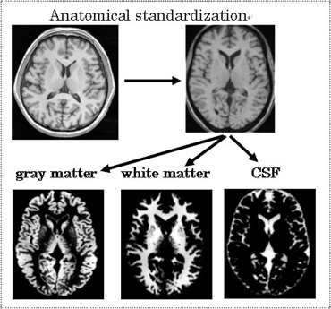
Fig. 3. Anatomical standardization and tissue segmenta-tion of the brain MRI
3.2. Voxel based image statistics
If gray matter probability in each voxel is considered as independent variable and subjects characteristics such as age, sex, blood pressure, blood glucose level to be as covariate, we can make voxel based image statistics. This analysis is named voxel based morphometry (VBM) and based on a general linear model in which each variable is independent and a linear summation of each. By this technique, we can detect brain regions in which gray matter volume correlate with some risk factors.
We also utilize deformation based morphomerty as an analytical tool. This technique provides a deformation vector to deform a brain to another brain, that is distances of two different brain in a space.
4. Establishment of Preventive Aging Medicine
The importance of research on aging is becoming more important for coming extremely aging society in Japan. In particular, prevention and early detection of age-related brain diseases, such as dementia is very important. In this project, we try to understand normal brain aging process and risk factors for it by using brain imaging analyses together with developing methodology for image processing and analyses. Final goal is to translate the results into clinical practice and to establish preventive aging medicine.
References
[1] Sato K, Taki Y, Fukuda H, and Kawashima R. Neuroanatomical database of normal Japanese brains. Neural Networks 16, 1301-1310, 2003.
[2] Taki Y, Goto R, Evans A, et al. Voxel-based morphometry of human brain with age and cerebrovascular risk factors. Neurobiol Aging 25, 455-63, 2004.
[3] Taki Y, Kinomura S, Sato K, et al. Both global gray matter volume and regional gray matter volume negatively correlate with lifetime alcohol intake in non-alcohol-dependent Japanese men: A volumetric analysis and a voxel-based morphometry. Alcohol Clin Exp Res 30, 1045-1050, 2006.
[4] Taki Y, Kinomura S, Awata S, et al. Male elderly subthreshold depression patients have smaller volume of medial part of prefrontal cortex and precentral gyrus compared with age-matched normal subjects: A voxel-based morphometry. J Affect Disord 88, 313-320, 2005.