HOME > Research Summaries > Nano-bio-imaging with Radiopharmaceuticals and Its Application to Health Sciences
Research Summaries

Nano-bio-imaging with Radiopharmaceuticals and Its Application to Health Sciences
Manabu Tashiro
Associate Professor
Division of Cyclotron Nuclear Medicine, Cyclotron and Radioisotope Center
E-mail:![]()
1. Introduction
The “tracer method” was first explored in the beginning of the 20th century by Dr. George von Hevesy, a Nobel laureate in chemistry in 1943, and later was combined with computer tomography technique in the end of the century. Currently the technique has been established as positron emission tomography (PET), that seems to be one of the most suitable tools for “nano-bio-imaging” since it can visualize pharmaco-dynamic and -kinetic information in the body of living subjects by injecting only a small amount of radiopharmaceuticals (just 1~several nmol).
Our group has been seeking further potentials and new applications of this radiological technique to clinical evaluation/diagnosis and to health promotion science. Our group is going to focus mainly the following subjects.
2. Research Projects
2.1. Establishment of models and simplified measurement protocols regarding nano-bio-imaging of neural functions in human brain
One of our main research topics is the elucidation of human brain functions. In the modern society, incidence of neuropsychiatric disorders such as cognitive disorder as well as metabolic disorders is constantly increasing as the mean age of the population increases. Various factors such as “stress” and “life style” have been regarded as among the main causes of the current situation of our society.
In human brain, neurotransmitters can manifest their effects with a very small amount. Therefore, it is not easy to visualize the actions of the neurotransmitters in the living human brain from outside without using a technique such as PET. With the PET, it is possible to quantify the interactions between transmitters and receptors as well as tissue metabolism, using the time course data of radioactivity in the blood and brain tissue. So far, our group has been studying the neuro-transmission of the histaminergic and dopaminergic neuronal systems by constructing a suitable model and a simplified quantification method that can be applied to clinical studies [1,2]. In addition, we are studying the side effects of centrally-acting drugs [3]. Currently, we are establishing a suitable pharmacokinetic model for quantification of beta-amyloid deposits in the brain of Alzheimer’s disease patients (Fig. 1), as well as modeling of neural transmission in the cholinergic neuronal system [4].
Development of new image processing methods is also important, such as more efficient software for co-registration of MRI and PET images (Fig. 1). We are working in this field of information science as well.
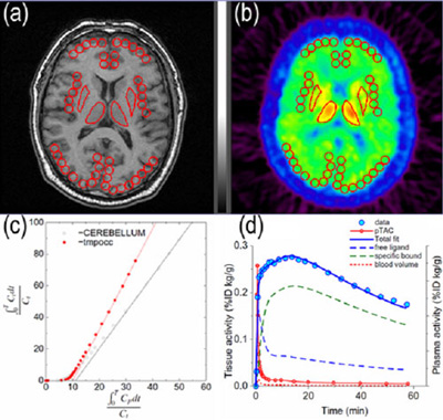
Fig. 1. Examples of pharmacokinetic analysis; regions of interest (ROI) defined in co-registered
(a) MRI and (b) PET images, (c) quantification based on graphical analysis and (d) compartment model analysis.
2.2. Application of preventive medicine and Health Promotion Sciences
According to the definition by the World Health Organization (WHO), “health” is “a complete state of physical, mental and social well-being, and not merely the absence of disease or infirmity.”. It is needless to say that the quality of life (QOL) is very important in all procedures of medical treatment and care. The definition may suggest that QOL in daily lives should be also paid attention as well.
In general, it has been thought, in Japan, anxiety toward radioactivity seems to be relatively stronger than in western countries. A highly-sensitive PET scanner has enabled examinations with minimal radiological doses in result with minimal radiation exposure far below the annual environmental exposure. Then, PET can be applied to elucidate the mechanism of our body functions to achieve and maintain healthy state. This could be termed as a new application to the field of preventive medicine and health promotion sciences.
So far, we have studied the brain activities of patients who suffer from malignant diseases. Our results have indicated that the brain metabolic pattern of cancer patients show abnormalities that correlate to the intensity of depressive mood. Our further studies have demonstrated a possible link between the psychological state and immune functions via the regional brain activities (Fig. 2) [5]. We have appealed that PET is useful not only for diagnosis of cancer but also for evaluation of psychological status of the patients who have cancer. Furthermore, we have conducted studies regarding whole-body exercise such as running and bicycle riding to observe regional activities of skeletal muscles [6], heart muscles, and brain [7,8]. PET seems to be useful to evaluate effects of various therapeutic interventions including exercise for metabolic disorders such as diabetes mellitus, hypertension, cerebro- and cardio-vascular diseases in terms of interaction between the mind, brain and body. Recently, alternative medicine has become popular among patients, and the scientific evaluation of these therapeutic techniques would be of clinical importance. PET can be very useful in evaluation based on scientific evidence.
We are going to observe the relationship between psycho-behavioral factors and achievement of healthy state in human body. And our ultimate goal is to establish the model of whole-body organ interactions.
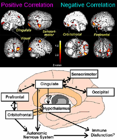
Fig. 2. Brain regions of positive and negative correlation to cellular immunity (Natural killer cell activity) in cancer patients (TOP); A hypothesized model of neuro-immune interaction in human (BOTTOM).
2.3. Development of High Resolution PET scanner specialized for B
In Japan, incidence and mortality from cancer has been increasing partly because of westernization of eating habit. For instance, the incidence of breast cancer has been constantly increasing among women.
Our group has started to develop positron emission mammography (PEM), a new PET system specialized for diagnosis of breast cancer, with excellent spatial resolution of 1 mm.
3. Conclusion
Thus, nano-bio-imaging technique using minimal-dose radiopharmaceuticals can have various potentials. For further development of this field, much deeper interaction of young researchers having a wide variety of academic backgrounds such as medical and pharmaceutical sciences, engineering, information science, etc. The author hopes that the present global COE program can supply an ideal environment for such fruitful interactions.
References
[1] Tashiro M, Duan X, Kato M, et al. Brain histamine H1 receptor occupancy of orally administered antihistamines, bepotastine and diphenhydramine, measured by PET with [11C]doxepin. Br J Clin Pharmacol 65, 811-821, 2008.
[2] Tashiro M, Mochizuki H, Sakurada Y, et al. Brain histamine H receptor occupancy of orally administered anti-histamines measured by positron emission tomography with [11C]doxepin in a placebo-controlled crossover study design in healthy subjects: a comparison of olopatadine and ketotifen. Br J Clin Pharmacol 61, 16-26, 2006.
[3] Tashiro M, Sakurada Y, Mochizuki H, et al. Effects of a sedative antihistamine, D-chlorpheniramine, on regional cerebral perfusion and performance during simulated car driving. Hum Psychopharmacol 23, 139-150, 2008.
[4] Kudo Y, Okamura N, Furumoto S, Tashiro M, et al. 2-(2-[2-Dimethyl aminothiazol-5-yl]ethenyl)-6-(2[fluoro] ethoxy) benzoxazole: a novel PET agent for in vivo detection of dense amyloid plaques in Alzheimer's disease patients. J Nucl Med 48, 553-561, 2007.
[5] Tashiro M, Itoh M, Kubota K, et al. Relationship between trait anxiety, brain activity and natural killer cell activity in cancer patients: a preliminary PET study. Psychooncology 10, 541-546, 2001.
[6] Fujimoto T, Itoh M, Tashiro M, et al. Glucose uptake by individual skeletal muscles during running using whole-body positron emission tomography. Eur J Appl Physiol 83, 297-302, 2000.
[7] Tashiro M, Itoh M, Fujimoto T, et al. Application of positron emission tomography to neuroimaging in sports sciences. Methods (in press).
[8] Tashiro M, Itoh M, Fujimoto T, et al. 18F-FDG PET mapping of regional brain activity in runners. J Sports Med Phys Fitness 41, 11-17, 2001.