HOME > Research Summaries > Development of Fully Implantable Retinal Prosthesis with 3-Dimensionally Stacked LSI
Research Summaries

Development of Fully Implantable Retinal Prosthesis with 3-Dimensionally Stacked LSI
Tetsu Tanaka
Professor
Department of Biomedical Engineering, Graduate School of Biomedical Engineering
E-mail:![]()
Abstract
We have proposed a novel retinal prosthesis with fully implantable 3-dimensionally (3-D) stacked retinal prosthesis chip. The retinal prosthesis chip consists of several LSI chips that are vertically stacked and electrically connected using 3D integration technology. Therefore, high fill factor, high resolution, and high sensitivity can be achieved in our retinal prosthesis.
To evaluate the optimal stimulating current to retinal cells, electrically evoked potential (EEP) was recorded in animal experiments using stimulating electrode array. The recorded EEP waveform shows a similar behavior to the visually evoked potential (VEP) waveform, indicating possibilities that the electrical stimulation of the retina can restore visual sensation for the blind patients. We will fabricate and implant the 3-D stacked retinal prosthesis chip for next 5 years.
1. Introduction
Recently, an increasing number of patients are suffering from retinitis pigmentosa (RP) and age-related macular degeneration (AMD). Several million patients become blind due to these diseases in the world. The RP and AMD result from impairment of photoreceptor cells that convert light signals to electrical signals. In actual, effective medical treatments for RP and AMD have not been established yet. While the photoreceptor cells degenerate with RP and AMD, many other retina cells (bipolar, horizontal, and ganglion) remain. Accordingly, it will be possible to recover one’s vision by stimulating the remaining retinal cells. Lots of research groups have proposed retinal prostheses to restore the visual sensation of blind patients [1]. The retinal prosthesis mainly consists of photodetectors, signal processing circuits, and stimulating electrode array. The photodetector receives optical signals and converts these into electrical signals. The processing circuits convert electrical signals to an appropriate pattern of electrical current. The stimulating electrode array placed on the retina stimulates remaining retinal cells. As results, the blind patients can perceive a dot of light at each stimulating point through an activation of their retinal cells.
We have been developing a fully implantable 3-D stacked retinal prosthesis chip using the 3-D integration technology [2, 3]. In our retinal prosthesis, all the parts of retinal prosthesis are implanted into an eyeball. By implanting all parts, blind patient can shift a gaze point by moving the eyeball and use own ocular system such as a lens and a cornea. Furthermore, we have been doing in-vivo experiments using the stimulating electrode array in order to optimize stimulating current.
2. Configuration of fully implantable retinal prosthesis
Figure 1 shows our retinal prosthesis with fully implantable 3-D stacked retinal prosthesis chip. The extraocular unit supplies electric power to the intraocular unit by electromagnetic induction. The intraocular unit consists of the 3-D stacked retinal prosthesis chip, a flexible printed circuit (FPC) with stimulating electrode array, and a secondary coil for receiving the power. In our retinal prosthesis, photodetectors in the top layer of 3-D stacked retinal prosthesis chip receive optical signals and convert them into image data. This image data is converted to appropriate patterns of electrical current by image processing circuits fabricated in the underlaid layer. Remaining retinal cells are activated by the electrical current through the stimulating electrode array placed on an inner retinal surface.
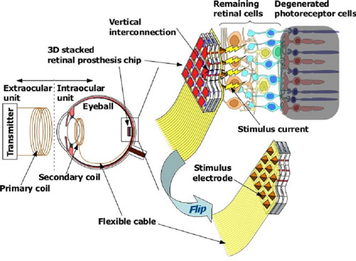
Fig. 1. Configuration of fully implantable retinal prosthesis with 3-D stacked LSI chip.
Figure 2 shows a SEM cross-section of 3-D stacked artificial retina chip fabricated for robot vision previously. Several circuit layers with different functions are stacked vertically and connected electrically by lots of vertical interconnections formed in the respective Si layers. This 3-D artificial retina chip provides a high speed, parallel processing capability, high fill factor, small form factor, and light weight. Our retinal prosthesis has many advantages similarly and it will provide a high quality of life (QOL) to the patients.
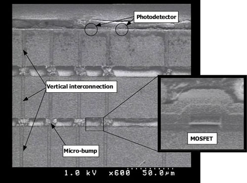
Fig. 2. SEM cross-section of artificial retina chip with 3-D structure.
3. EEP recording using stimulating electrode array on polyimide-based FPC
The retina of blind patients has to be stimulated with optimal stimulating current to obtain proper visual information. To optimize stimulating current parameters, it is very important to record the EEP, which indicates electrical activities in the primary visual cortex. Stimulating current parameters can be optimized by comparing EEP with VEP.
We fabricated the implantable Al stimulating electrode array on polyimide-based FPC. The FPC has a length of 140 mm, a width of 2 mm, and a thickness of 30 μm. At the end of the FPC, 4×4 stimulating electrodes are formed. The stimulating electrodes are 70 μm in diameter and are placed at a pitch of 200 μm. At the other end of the FPC, 16 pads for connection to a stimulating current generator are formed. Figure 3 shows the fabricated stimulating electrode array.
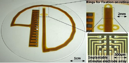
Fig. 3. Photographs of stimulating electrode array on polyimide-based FPC.
We measured the EEP when the retina of a rabbit was stimulated with implanted stimulating electrode array. In this experiment, Japanese white rabbits (2-3kg) were used. All procedures adhered to the Association for Research in Vision and Ophthalmology (ARVO) Resolution on the Use of Animals in Research. The stimulating electrode array was fixed on the surface of the retina by a retinal tack. The EEP response was successfully recorded, and the EEP waveform shows a similar behavior to the visually evoked potential (VEP) waveform. Figure 4 shows relationships between the amplitude of recorded EEP and the electrical charge of stimulating current. Positive relationships can be found between quantity of electrical charges and amplitude of EEP, which results in the increase of the number of activated retinal cells.
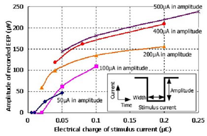
Fig. 4. Relationships between EEP amplitude and electrical charge of stimulus current.
4. Conclusion
We proposed the novel retinal prosthesis with fully implantable 3-D stacked LSI chip. Our retinal prosthesis has many advantages such as high resolution, small size, and light weight, and give patients high QOL. We compared EEP to VEP in order to optimize the stimulating current parameters. The recorded EEP waveform showed a similar behavior to VEP waveform, which indicates a great expectation for restoring the visual sensation by stimulating current to retinal cells.
We will fabricate and implant the 3-D stacked retinal prosthesis chip for next 5 years.
Acknowledgement
T. Tanaka acknowledges the support of Tohoku University Global COE Program “Global Nano-Biomedical Engineering Education and Research Network Centre”.
References
[1] Humayun MS, de Juan E, Weiland JD, Dagnelie G, Katona S, Greenberg R, Suzuki S. Pattern electrical stimulation of the human retina. Vision Research 39, 2569-2576, 1999.
[2] Kurino H, Nakagawa Y, Nakamura T, Yamada Y, Lee K, Koyanagi M. Biologically Inspired Vision Chip with Three Dimensional Structure. The Institute of Electronics, Information and Communication Engineers Transactions on Electronics E84-C, 1717-1722, 2001.
[3] Koyanagi M, Nakagawa Y, Lee K, Nakamura T, Yamada Y, Inamura K, Park K, Kurino K. Neuromorphic Vision Chip Fabricated Using Three-Dimensional Integration Technology. In Proceedings of IEEE International Solid State Circuits Conference, San Francisco, 270-271, 2001.