HOME > Research Summaries > Study on Ultrasonic Elasticity Imaging for Arterial Tissue Characterization
Research Summaries

Study on Ultrasonic Elasticity Imaging for Arterial Tissue Characterization
Hiroshi Kanai* and Hideyuki Hasegawa
Professor
Department of Electronic Engineering, Graduate School of Engineering
E-mail:![]()
Abstract
Knowledge of the physical properties of atherosclerotic plaque is essential when evaluating its vulnerability in a clinical setting. Such knowledge, however, is still difficult to obtain with the various approaches developed to date. We have developed a noninvasive method for evaluating the regional elasticity (the elastic modulus in the circumferential direction) of tissue surrounding atherosclerotic plaque in which a novel approach is applied to measure minute changes in thickness of each of the multiple layers of the arterial wall during one heartbeat. By comparing the pathological findings with the distribution of elasticity, average elasticity of lipid and that of a mixture of smooth muscle and collagen fiber can be determined. Based on these reference parameters, each point is statistically categorized as lipid, mixture, or other. Thus, the plaque is electronically stained using transcutaneous ultrasound. This novel method offers potential as a diagnostic technique for detection of plaque vulnerability with high spatial resolution.
1. Introduction
The steady increase in the number of patients with myocardial infarction or cerebral infarction, both of which are considered to be mainly caused by atherosclerosis, is becoming a serious problem. Therefore, it is important to diagnose atherosclerosis in the early stage. Computed tomography (CT), magnetic resonance imaging (MRI) and ultrasonic diagnostic equipment are employed for the diagnosis of atherosclerosis.
However, they provide information only on the shape of the artery such as the diameter of the lumen. In addition, CT and MRI are not suitable for repetitive diagnosis due to the exposure to radiation and the high expense. However, the diameter of the lumen, which can be measured by these methods, is not changed by early-stage atherosclerosis. In addition, it is essential to evaluate the mechanical properties and tissue composition of atherosclerotic plaque for diagnosing its vulnerability. Therefore, it is necessary to develop a method for measuring the elastic property of the arterial wall..
2. Conventional Measurements of Elasticity
To measure the elasticity of the arterial wall, the pulse wave velocity (PWV) method was developed as a technique for the noninvasive diagnosis of atherosclerosis [1]. In this method, the elasticity of the arterial wall is evaluated by measuring the velocity of the pressure wave propagating from the heart to the femoral artery. Though it is useful in terms of noninvasive evaluation of elasticity, regional properties cannot be evaluated due to a low spatial resolution of tens of centimeters, which almost corresponds to the distance from the heart to the femoral artery.
Methods for measurement of the change in artery diameter have been proposed. in order to obtain the circumferential distensibility of the arterial wall in the plane which is perpendicular to the axial direction of the artery. By assuming the artery to be a cylindrical shell, the average elasticity of the entire circumference of the plane can be evaluated [2,3]. However, the regional elasticity of atherosclerotic plaque cannot be obtained by these methods, because an artery with such plaque cannot be assumed to be a cylindrical shell with uniform wall thickness.
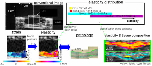
Fig. 1. Tissue classification of arterial wall by noninvasive elasticity measurement.
3. Exploration of High Resolution Ultrasound Elasticity Imaging
In our series of studies, we have been attempting to evaluate the elasticity of a local region, even in the case of atherosclerotic plaque using transcutaneous ultrasound (Fig.1) [4-6]. Such a technique for noninvasive measurement of the spatial distribution of the regional elasticity would be useful for diagnosis of the vulnerability of such plaque as well as for the diagnosis of early-stage atherosclerosis. In our method, the small displacement gradient (change in thickness) of the arterial wall due to the heartbeat was accurately measured in each local region which corresponds to the focal area of the ultrasonic beam. From the resultant displacement gradient, the regional strain and the elasticity of the arterial wall were noninvasively evaluated. Recently many studies were conducted to measure the elasticity of the arterial wall with ultrasound [7-9].
In addition to diagnosis of developed atherosclerosis, it is also important to diagnose atherosclerosis in its early stage. Ross hypothesized that endothelium cells, which covers the luminal surface of the arterial wall (Fig. 2), are injured in the early-stage atherosclerosis [10]. The endothelial function can be evaluated by measuring the change in elasticity of the arterial wall due to the endothelial-dependent dilation after avascularization because the endothelium cells produces nitric oxide (NO) in response to the increased shear stress due to recirculation and NO produced by endothelium cells relaxes smooth muscle in the media.
As described above, ultrasound elasticity imaging of the arterial wall has various possibilities for accurate diagnosis of atherosclerosis.
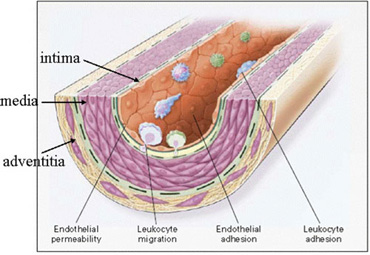
Fig. 2. Structure of arterial wall [10]
4. Conclusion
High-resolution images of arterial walls can be obtained by recent ultrasonic diagnostic equipment. However, morphological information is mainly derived from such cross-sectional ultrasound images whereas the mechanical properties and tissue composition in the arterial wall are more important for diagnosis of atherosclerosis. Ultrasound elasticity imaging based on the detailed analysis of ultrasound signal provides additional useful information for accurate diagnosis of both developed and early-stage atherosclerosis.
References
[1] Hallock P. Arterial elasticity in man in relation to age as evaluated by the pulse wave velocity method. Arch Int Med 54, 770-798, 1934.
[2] Hoeks APG, Ruissen CJ, Hick P, and Reneman RS. Transcutaneous detection of relative changes in artery diameter. Ultrasound Med Biol 11, 51-59, 1985.
[3] Hayashi K, Handa H, Nagasawa S, Okumura A, and Moritake K. Stiffness and elastic behavior of human intracranial and extracranial arteries. J Biomech 13, 175-184, 1980.
[4] Kanai H, Sato M, Chubachi N, and Koiwa Y. Transcutaneous measurement and spectrum analysis of heart wall vibrations. IEEE Trans Ultrason Ferroelectr Freq Control 43, 791-810, 1996.
[5] Hasegawa H, Kanai H, Hoshimiya N, and Koiwa Y. Evaluating the regional elastic modulus of a cylindrical shell with nonuniform wall thickness. J Med Ultrason 31, 81-90, 2004.
[6] Kanai H, Hasegawa H, Ichiki M, Tezuka F, and Koiwa Y. Elasticity imaging of atheroma with transcutaneous ultrasound -preliminary study-. Circulation 107, 3018-3021, 2003.
[7] de Korte CL, C市pedes, van der Steen AFW, and Lanc仔. Intravascular elasticity imaging using ultrasound: Feasibility studies in phantoms. Ultrasound Med Biol 23, 735-746, 1997.
[8] Rabben SI, Bjセrum S, Sソrhus, and Torp H. Ultrasound-based vessel wall tracking: An auto-correlation technique with RF center frequency estimation. Ultrasound Med Biol 28, 507-517, 2002.
[9] Maurice RL, Ohayon J, Fr師igny Y, Bertrand M, Soulez G, and Cloutier G. Noninvasive vascular elastography: Theoretical framework. IEEE Trans Med Imaging 23, 164-180, 2004.
[10] Ross R. Atherosclerosis -an inflammatory disease-. New Engl J Med 340, 115-126, 1999.